Table of contents
- Learning Objectives
- How acid–base balance works
- Step 1: Metabolism continuously produces acid
- Step 2: Buffers limit the immediate pH change
- Step 3: The lungs regulate carbon dioxide within minutes
- Step 4: The kidneys preserve bicarbonate and excrete nonvolatile acid
- Step 5: pH depends on the relationship between bicarbonate and carbon dioxide
- Step 6: Primary disorders arise when one control system fails or is driven abnormally
- What the blood gas actually reports
- ABG or VBG?
- Practice: explain the physiology before opening the answer
- Summary
- References
Critical Care · Acid Base
Acid-Base Physiology
Acid–Base Physiology: Buffers, Lungs, and Kidneys
Learning Objectives
By the end of this lesson, you should be able to:
- Explain how metabolism produces volatile and nonvolatile acids.
- Describe how bicarbonate, hemoglobin, proteins, and phosphate limit rapid changes in pH.
- Explain how ventilation changes carbon dioxide and pH within minutes.
- Explain how the kidneys reclaim bicarbonate, generate new bicarbonate, and excrete acid.
- Distinguish acidemia and alkalemia from the processes called acidosis and alkalosis.
- Explain what each blood-gas value represents and when an arterial sample is preferable to a venous sample.
How acid–base balance works
Normal arterial pH is approximately 7.35–7.45, although laboratory reference intervals vary. That narrow range is maintained through a sequence of four processes: the body produces acid, buffers limit the immediate change in pH, the lungs regulate carbon dioxide, and the kidneys regulate bicarbonate and eliminate nonvolatile acid.
Step 1: Metabolism continuously produces acid
Cells generate two broad categories of acid:
- Volatile acid: Aerobic metabolism produces carbon dioxide (CO₂). In water, CO₂ combines with water to form carbonic acid, which can dissociate into hydrogen ions (H⁺) and bicarbonate (HCO₃⁻). Because CO₂ can be exhaled, the lungs can eliminate this acid load.
- Nonvolatile or fixed acid: Metabolism of sulfur-containing amino acids and phosphates generates acids that cannot be exhaled. Lactate, ketoacids, and ingested toxins can add much larger acid loads during illness. These acids must ultimately be metabolized or excreted by the kidneys.
The central reversible reaction is:
CO₂ + H₂O ⇌ H₂CO₃ ⇌ H⁺ + HCO₃⁻
The enzyme carbonic anhydrase rapidly accelerates this reaction in red blood cells, renal tubular cells, and other tissues. The reaction can move in either direction. Adding CO₂ pushes it toward more hydrogen ions; removing CO₂ pulls it toward less hydrogen ion.
Step 2: Buffers limit the immediate pH change
When hydrogen ions appear, buffers bind some of them within seconds. The bicarbonate system is the principal extracellular buffer, while hemoglobin, intracellular proteins, and phosphate also contribute.
For example, when a nonvolatile acid releases H⁺, bicarbonate binds that H⁺ and is converted through carbonic acid into CO₂ and water. The lungs then exhale the generated CO₂. The blood bicarbonate concentration falls because bicarbonate was consumed while buffering the acid.
This explains an important laboratory pattern: in metabolic acidosis, a low bicarbonate is not merely a marker. It reflects bicarbonate consumption, bicarbonate loss, or impaired renal bicarbonate generation.
Step 3: The lungs regulate carbon dioxide within minutes
Carbon dioxide production and alveolar ventilation determine arterial carbon dioxide tension (PaCO₂):
- If alveolar ventilation increases, more CO₂ is exhaled, PaCO₂ falls, the reaction shifts left, and pH rises.
- If alveolar ventilation decreases, CO₂ accumulates, PaCO₂ rises, the reaction shifts right, and pH falls.
This is why primary respiratory disorders are defined by their carbon dioxide change:
- Respiratory acidosis: inadequate effective ventilation causes CO₂ retention.
- Respiratory alkalosis: ventilation exceeds CO₂ production, causing excessive CO₂ removal.
Chemoreceptors respond to changes in carbon dioxide, pH, and oxygenation. In metabolic acidosis, increased ventilation can lower PaCO₂ within minutes. This response reduces the fall in pH but cannot replace bicarbonate or remove the original nonvolatile acid.
Step 4: The kidneys preserve bicarbonate and excrete nonvolatile acid
The kidneys perform three related tasks:
- Reclaim filtered bicarbonate. Nearly all filtered bicarbonate is normally returned to the blood, primarily in the proximal tubule. Failure to reclaim it causes urinary bicarbonate loss.
- Excrete hydrogen ions as titratable acid. Secreted H⁺ binds urinary buffers, especially phosphate, allowing acid to leave the body without lowering urine pH indefinitely.
- Generate and excrete ammonium. Proximal tubular metabolism of glutamine produces ammonium (NH₄⁺) and new bicarbonate. Ammonium is ultimately trapped in the urine and excreted, while the new bicarbonate enters the blood. This pathway can increase substantially during sustained acidosis.
The kidney response takes hours to begin and several days to reach its full effect. In chronic respiratory acidosis, persistently elevated CO₂ stimulates greater bicarbonate retention and net acid excretion. In chronic respiratory alkalosis, the kidneys excrete more bicarbonate. Kidney failure, hypoaldosteronism, tubular disease, or severe hyperkalemia can impair these responses.
Step 5: pH depends on the relationship between bicarbonate and carbon dioxide
The Henderson–Hasselbalch relationship can be written as:
pH = 6.1 + log[HCO₃⁻ / (0.03 × PaCO₂)]
You do not need to calculate this equation at the bedside. Its value is conceptual: pH depends on the ratio of bicarbonate to dissolved carbon dioxide, not on either value alone.
- A fall in bicarbonate lowers the ratio and lowers pH unless ventilation also lowers CO₂.
- A rise in carbon dioxide lowers the ratio and lowers pH unless the kidneys retain more bicarbonate.
- A rise in bicarbonate or fall in carbon dioxide raises the ratio and raises pH.
- If bicarbonate and carbon dioxide change in the same direction and in the expected proportion, one is usually the primary disturbance and the other is compensation.
- If they change by an unexpected amount—or move in directions that cannot be explained by compensation—more than one primary disorder is present.
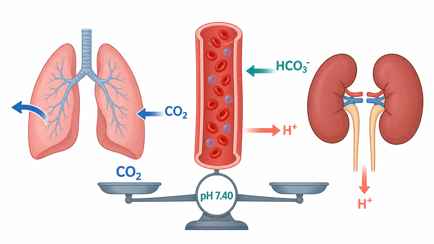
A low pH is acidemia; a high pH is alkalemia. An acidosis is a process that pushes pH down, whereas an alkalosis pushes it up. These terms are not interchangeable. A patient can have metabolic acidosis and respiratory alkalosis at the same time, leaving the measured pH near the reference interval. Therefore, never stop after deciding that the pH is “normal.”
Step 6: Primary disorders arise when one control system fails or is driven abnormally
| Primary disorder | Initial physiologic problem | Immediate effect | Expected compensation |
|---|---|---|---|
| Metabolic acidosis | Acid is added, bicarbonate is lost, or renal acid excretion fails | HCO₃⁻ falls and pH falls | Ventilation increases, lowering PaCO₂ |
| Metabolic alkalosis | Hydrogen ions are lost or bicarbonate is gained and retained | HCO₃⁻ rises and pH rises | Ventilation decreases modestly, raising PaCO₂ |
| Respiratory acidosis | Alveolar ventilation is inadequate | PaCO₂ rises and pH falls | Kidneys retain/generate HCO₃⁻ and excrete more acid |
| Respiratory alkalosis | Alveolar ventilation is excessive for CO₂ production | PaCO₂ falls and pH rises | Kidneys excrete more HCO₃⁻ and reduce net acid excretion |
Compensation is limited. Hypoventilation during metabolic alkalosis is constrained by the need to maintain oxygenation. Respiratory compensation for metabolic acidosis is constrained by respiratory muscle capacity and lung disease. Renal compensation is limited by kidney function, distal sodium delivery, potassium balance, and the time available for adaptation.
What the blood gas actually reports
| Value | What it represents | Typical arterial reference | Key caution |
|---|---|---|---|
| pH | Net acid–base state | 7.35–7.45 | A near-normal value can conceal mixed disease. |
| PaCO₂ | Arterial partial pressure of CO₂ | 35–45 mmHg | Primarily reflects alveolar ventilation. |
| HCO₃⁻ | Calculated bicarbonate on most analyzers | 22–26 mmol/L | May differ slightly from chemistry total CO₂. |
| Base excess | About −2 to +2 mmol/L | Helpful adjunct, not a replacement for the full analysis. | |
| PaO₂ | Arterial partial pressure of oxygen | Context dependent | Interpret with inspired oxygen, age, altitude, and clinical setting. |
The serum chemistry bicarbonate is usually reported as total carbon dioxide. The blood-gas bicarbonate is commonly calculated from pH and measured carbon dioxide. Small differences are expected; large differences should prompt review of sampling time, processing, and specimen identity.
Preanalytical problems can create false patterns:
- Air bubbles tend to move the sample toward room-air gas values.
- Delayed analysis allows ongoing cellular metabolism to consume oxygen and generate carbon dioxide and acid.
- Excess liquid heparin can dilute the specimen.
- A sample drawn while ventilation or perfusion is rapidly changing may become obsolete within minutes.
- A mislabeled venous sample can mimic severe arterial hypoxemia.
ABG or VBG?
A peripheral VBG is often sufficient for an initial acid–base assessment in a hemodynamically stable adult. Venous pH usually tracks arterial pH closely enough to identify clinically important acidemia or alkalemia. However:
- Venous carbon dioxide is not an exact substitute for arterial carbon dioxide.
- Venous oxygen tension must not be used to assess arterial oxygenation.
- Pulse oximetry estimates saturation but does not provide pH, ventilation, or a reliable value in every dyshemoglobinemia or low-perfusion state.
Choose an ABG when you need precise arterial oxygenation, an exact arterial carbon dioxide value, assessment of severe or changing respiratory failure, or clarification when the VBG and bedside picture disagree. Use co-oximetry when carbon monoxide exposure, methemoglobinemia, or another dyshemoglobinemia is suspected.
Practice: explain the physiology before opening the answer
- Why does bicarbonate fall when a nonvolatile acid is added to the blood?
- Why can the lungs compensate for metabolic acidosis but not eliminate its cause?
- What is the difference between reclaiming filtered bicarbonate and generating new bicarbonate?
- Why can a patient have a near-normal pH despite two serious acid–base disorders?
- Which measurements from a peripheral venous blood gas should not be treated as exact substitutes for arterial carbon dioxide or oxygen tension?
Show physiology answers
- Bicarbonate accepts hydrogen ions and is converted through carbonic acid into carbon dioxide and water. The bicarbonate concentration therefore falls as it buffers the added acid.
- Increased ventilation removes carbon dioxide and raises the bicarbonate-to-carbon-dioxide ratio, limiting the fall in pH. It does not metabolize or excrete the original lactate, ketoacid, toxin, or other fixed acid.
- Reclamation prevents filtered bicarbonate from being lost in urine. New bicarbonate is added to the extracellular fluid when the kidney excretes hydrogen ions as ammonium or titratable acid.
- Opposing primary processes can move pH in opposite directions. For example, metabolic acidosis and respiratory alkalosis may coexist and produce a pH near the reference interval.
- Venous carbon dioxide is not an exact arterial substitute, and venous oxygen tension must not be used to assess arterial oxygenation.
Summary
- Metabolism continuously produces carbon dioxide and nonvolatile acids.
- Buffers limit immediate changes in free hydrogen-ion concentration but do not eliminate the underlying acid load.
- The lungs regulate carbon dioxide within minutes by changing alveolar ventilation.
- The kidneys preserve filtered bicarbonate, generate new bicarbonate, and excrete nonvolatile acid over hours to days.
- pH depends on the relationship between bicarbonate and dissolved carbon dioxide.
- Acidemia and alkalemia describe the measured pH; acidosis and alkalosis describe processes that push pH down or up.
- A venous blood gas often supports an initial acid–base assessment, but arterial sampling is needed when precise arterial oxygenation or carbon dioxide measurement changes the decision.
References
- Adrogué HJ, Gennari FJ, Galla JH, Madias NE. Assessing acid-base disorders. Kidney International. 2009. Link
- Kraut JA, Madias NE. Metabolic acidosis: pathophysiology, diagnosis and management. Nature Reviews Nephrology. 2010. Link
- Byrne AL, Bennett M, Chatterji R, et al. Peripheral venous and arterial blood gas analysis in adults: are they comparable? A systematic review and meta-analysis. Respirology. 2014. Link
- Weimar Z, Smallwood N, Shao J, et al. Arterial blood gas analysis or venous blood gas analysis for adult hospitalised patients with respiratory presentations: a systematic review. Internal Medicine Journal. 2024. Link
Last Edited 07/17/2026