Table of contents
- Learning Objectives
- Brief physiology recap
- The five-step interpretation sequence
- Step 1: Name the pH state
- Step 2: Identify the primary direction
- Step 3: Test compensation
- Step 4: Calculate the anion gap
- Step 5: Return to the patient
- Metabolic acidosis: first divide by anion gap
- High-anion-gap metabolic acidosis
- Normal-anion-gap metabolic acidosis
- Metabolic alkalosis: generation plus maintenance
- Respiratory acidosis: too little effective ventilation
- Respiratory alkalosis: ask why the patient is hyperventilating
- Mixed disorders: the normal pH trap
- Practice: interpret before opening the answer
- Summary
- References
Critical Care · Acid Base
Acid Base Diagnosis
Acid–Base Disorders: Stepwise Blood Gas Interpretation
Learning Objectives
By the end of this lesson, you should be able to:
- Interpret an arterial blood gas or venous blood gas using a consistent five-step sequence.
- Identify the primary acid–base disorder and test whether compensation is appropriate.
- Calculate the anion gap, correct it for albumin, and use the delta ratio to uncover additional metabolic disorders.
- Build focused differentials for high-gap and normal-gap metabolic acidosis.
- Distinguish major causes of metabolic alkalosis using clinical context and urine chloride.
- Distinguish acute from chronic respiratory acidosis and respiratory alkalosis.
- Recognize common mixed acid–base disorders, including patterns with a near-normal pH.
- Apply the full interpretation sequence to clinical cases.
Brief physiology recap
Blood pH depends on the relationship between bicarbonate and dissolved carbon dioxide. Metabolic disorders begin with a change in bicarbonate or fixed-acid balance; respiratory disorders begin with a change in carbon dioxide caused by ventilation. Compensation changes the other component in the direction that limits the pH disturbance, but it does not remove the primary cause. A response outside the expected range indicates an additional primary disorder.
The five-step interpretation sequence
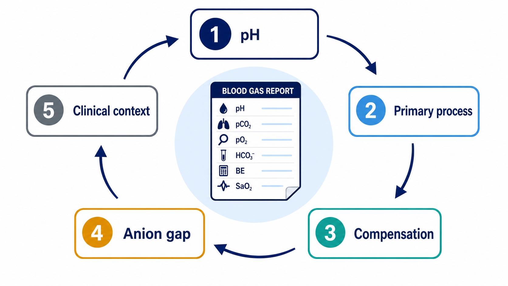
Step 1: Name the pH state
- pH below the reference interval: acidemia.
- pH above the reference interval: alkalemia.
- pH within the interval: still inspect both carbon dioxide and bicarbonate for opposing disorders.
Step 2: Identify the primary direction
| Primary process | pH direction | Primary change |
|---|---|---|
| Metabolic acidosis | ↓ | HCO₃⁻ ↓ |
| Metabolic alkalosis | ↑ | HCO₃⁻ ↑ |
| Respiratory acidosis | ↓ | PaCO₂ ↑ |
| Respiratory alkalosis | ↑ | PaCO₂ ↓ |
If both carbon dioxide and bicarbonate appear to explain the pH, decide which change is primary using the clinical setting and the expected compensatory response.
Step 3: Test compensation
| Primary disorder | Expected response |
|---|---|
| Metabolic acidosis | Expected PaCO₂ ≈ 1.5 × HCO₃⁻ + 8 ± 2 |
| Metabolic alkalosis | PaCO₂ rises about 0.7 mmHg per 1 mmol/L rise in HCO₃⁻; usually does not exceed about 55 mmHg from compensation alone |
| Acute respiratory acidosis | HCO₃⁻ rises about 1 mmol/L per 10 mmHg PaCO₂ rise |
| Chronic respiratory acidosis | HCO₃⁻ rises about 4 mmol/L per 10 mmHg PaCO₂ rise |
| Acute respiratory alkalosis | HCO₃⁻ falls about 2 mmol/L per 10 mmHg PaCO₂ fall |
| Chronic respiratory alkalosis | HCO₃⁻ falls about 4 mmol/L per 10 mmHg PaCO₂ fall |
These are approximations, not biological laws. Extreme values, evolving illness, kidney dysfunction, treatment, and laboratory variation reduce precision.
Worked compensation example
A patient has pH 7.22, HCO₃⁻ 12 mmol/L, and PaCO₂ 26 mmHg.
Expected PaCO₂ = 1.5 × 12 + 8 ± 2 = 26 ± 2 mmHg.
The measured PaCO₂ is within the expected range, so this is a metabolic acidosis with appropriate respiratory compensation. If PaCO₂ were 40 mmHg, an additional respiratory acidosis would be present. If it were 18 mmHg, an additional respiratory alkalosis would be present.
Why an elevated baseline bicarbonate matters
A patient with stable chronic obstructive pulmonary disease (COPD) has a baseline pH of 7.36, PaCO₂ of 60 mmHg, and HCO₃⁻ of 33 mmol/L. The high bicarbonate is expected renal compensation for chronic respiratory acidosis; it is not a primary metabolic alkalosis by itself.
The patient later develops profuse diarrhea. The new gas shows pH 7.28, PaCO₂ 60 mmHg, and HCO₃⁻ 27 mmol/L. A bicarbonate of 27 mmol/L is still above the usual reference interval, but it has fallen substantially below this patient's compensated baseline. The correct interpretation is chronic respiratory acidosis plus a new metabolic acidosis, not chronic respiratory acidosis with “normal” renal compensation.
Baseline values matter because a result can be numerically high yet pathologically low for that patient. The same principle works in the opposite direction: a bicarbonate substantially above the chronic baseline suggests an additional metabolic alkalosis, such as from loop diuretics, vomiting, or post-hypercapnic physiology.
Step 4: Calculate the anion gap
Anion gap = sodium − (chloride + bicarbonate)
Use your laboratory's reference interval. Potassium is usually omitted. Because albumin is a major unmeasured anion, hypoalbuminemia can conceal a clinically important gap:
Corrected gap ≈ measured gap + 2.5 × (4.0 − albumin in g/dL)
Why calculate the gap when acidosis is not primary?
The anion gap is a screen for accumulated unmeasured anions, not merely a label for the primary pH disorder.
- Lactate, ketoanions, toxic alcohol metabolites, retained acids in kidney failure, and other anions increase the gap as they accumulate.
- A simultaneous respiratory alkalosis or metabolic alkalosis may raise the pH or bicarbonate enough to hide the expected acidemia.
- Hypoalbuminemia may lower the measured gap and further conceal the rise.
- Therefore, calculate and albumin-correct the gap during every complete blood-gas interpretation when the necessary chemistry values are available—even when alkalemia, respiratory disease, or a near-normal pH appears to be the primary pattern.
An elevated corrected gap then triggers a search for the accumulated anion and a delta-ratio calculation. A normal gap does not exclude every serious illness, but skipping the calculation can miss a clinically important mixed disorder.
Step 5: Return to the patient
Ask what process can produce this pattern now. Review vital signs, ventilation, perfusion, renal function, glucose and ketones, lactate, medications, losses, toxic exposures, and treatment already given. Repeat the gas when physiology is changing rapidly.
Metabolic acidosis: first divide by anion gap
Metabolic acidosis is a fall in bicarbonate caused by added acid, impaired renal acid excretion, or bicarbonate loss. Respiratory compensation lowers carbon dioxide; inadequate compensation can rapidly worsen acidemia.
High-anion-gap metabolic acidosis
For every elevated albumin-corrected anion gap, complete the following sequence:
- Confirm the chemistry values were obtained at approximately the same time and compare with the laboratory's reference gap.
- Identify the likely unmeasured anion using the clinical setting and targeted tests.
- Calculate the delta ratio to look for another metabolic disorder.
- If toxic alcohol or another osmotically active ingestion is plausible, calculate the serum osmolal gap from a contemporaneous sample and obtain targeted concentrations. Do not order or interpret the osmolal gap as a universal high-gap screen.
| Mechanism | Important causes | Useful clues and tests |
|---|---|---|
| Lactate accumulation | Shock, sepsis, severe hypoxemia, seizures, regional ischemia, selected drugs or toxins | Perfusion assessment, serial lactate, source evaluation |
| Ketoacid accumulation | Diabetic, alcoholic, starvation, pregnancy-associated, sodium-glucose cotransporter-2 inhibitor–associated | Serum beta-hydroxybutyrate, glucose, medication and nutrition history |
| Reduced renal acid excretion | Advanced kidney failure | Creatinine trend, urine output, potassium, clinical volume status |
| Toxic alcohol metabolites | Methanol, ethylene glycol | Exposure history, osmolal gap, targeted levels, visual or renal findings |
| Salicylate | Acute or chronic poisoning | Serial salicylate levels, tinnitus, tachypnea, temperature, mental status |
| Other acids | Selected medications and uncommon metabolic disorders | Medication, nutrition, and exposure review |
Delta ratio: calculation and interpretation
Calculate the delta ratio in every patient with an elevated corrected anion gap. It compares the rise in anion gap with the fall in bicarbonate:
Delta ratio = (anion gap − normal gap) / (24 − HCO₃⁻)
Use the albumin-corrected gap and the local normal gap when available. If the denominator is zero or negative because bicarbonate is 24 mmol/L or higher, the ratio is not mathematically useful; the elevated gap itself still requires explanation.
| Delta ratio | Practical interpretation |
|---|---|
| <1 | The bicarbonate fell more than the gap rose: high-gap acidosis plus an additional normal-gap metabolic acidosis is likely |
| Approximately 1–2 | The rise in gap and fall in bicarbonate are broadly proportional: a predominantly high-gap metabolic acidosis is likely |
| >2 | The bicarbonate fell less than the gap rose: suspect an additional metabolic alkalosis or a high pre-illness bicarbonate, including compensated chronic respiratory acidosis |
These ranges are guides rather than rigid diagnostic boundaries. Baseline anion gap and bicarbonate, albumin correction, kidney function, timing, saline administration, urinary ketoanion loss, and a resolving process can shift the ratio.
Delta ratio physiology, step by step
- An added nonchloride acid dissociates into hydrogen ions and an accompanying anion, such as lactate or a ketoanion.
- Bicarbonate buffers the hydrogen ions, so serum bicarbonate falls.
- The accompanying anion remains in the extracellular fluid and raises the anion gap.
- In an uncomplicated high-gap acidosis, the rise in gap and fall in bicarbonate are therefore roughly proportional.
- If bicarbonate falls more than the gap rises, another process is consuming or removing bicarbonate without adding an unmeasured serum anion. Diarrhea, renal tubular acidosis, or chloride-rich fluid can produce this additional normal-gap acidosis.
- If bicarbonate falls less than the gap rises, another process is raising bicarbonate or the patient started with an elevated bicarbonate. Vomiting, diuretics, contraction alkalosis, or chronic hypercapnic compensation are common explanations.
The delta ratio does not identify the cause by itself. It tells you that the observed bicarbonate cannot be explained by the high-gap process alone and directs the next clinical question.
Osmolal gap: when and how to use it
Use the osmolal gap when the history or presentation raises concern for methanol, ethylene glycol, isopropanol, propylene glycol, diethylene glycol, or another osmotically active exposure. Relevant clues include unexplained high-gap acidosis, visual symptoms, acute kidney injury or calcium oxalate crystalluria, central nervous system depression, an exposure history, or an unexplained measured hyperosmolality.
- Obtain measured serum osmolality, sodium, glucose, blood urea nitrogen (BUN), and ethanol from the same time point.
- In conventional US units, one common estimate is: calculated osmolality = 2 × sodium + glucose/18 + BUN/2.8. Account for ethanol using the laboratory's validated method.
- Osmolal gap = measured osmolality − calculated osmolality. The equation and local reference interval matter; different accepted equations produce different gaps.
- A markedly elevated result—for example, approximately 20 mOsm/kg or more—strengthens concern for an unmeasured osmole, but it is not specific for a toxic alcohol. Ethanol, ketoacidosis, kidney failure, shock, mannitol, contrast agents, and laboratory variation may contribute.
- A normal result does not exclude toxic alcohol poisoning. Early after ingestion, unmetabolized parent alcohol may produce a large osmolal gap before much acidosis develops. Later, conversion to acidic metabolites can produce a large anion gap after the osmolal gap has declined.
Send targeted toxic alcohol concentrations when available and contact a poison center or medical toxicologist promptly. Do not delay time-sensitive treatment solely while waiting for a gap or confirmatory level.
Review toxic alcohol diagnosis · Review osmolal-gap limitations
Normal-anion-gap metabolic acidosis
Here, lost bicarbonate or impaired renal acid excretion is largely replaced by chloride, producing hyperchloremic acidosis.
When direct urine ammonium is unavailable, obtain a contemporaneous urine sodium, potassium, and chloride and calculate:
Urine anion gap = urine sodium + urine potassium − urine chloride
Use the result only if urine collection and distal sodium delivery are adequate and no major interfering urinary anion is present.
| Urine anion gap pattern | Physiologic response | Differential diagnosis |
|---|---|---|
| Clearly negative | Urinary ammonium chloride excretion is increased; the kidney is responding to systemic acidosis | Gastrointestinal bicarbonate loss from diarrhea, fistula, or high-output drainage; chloride-load acidosis with preserved renal response; some cases of proximal renal tubular acidosis once serum bicarbonate is below the reduced reabsorptive threshold |
| Near zero or positive | Ammonium excretion is inappropriately low or not detectably increased | Distal renal tubular acidosis (type 1), type 4 renal tubular acidosis, reduced ammonium excretion in kidney disease, or inadequate distal sodium delivery |
Then use potassium, urine pH, kidney function, medication history, and the clinical setting to refine the diagnosis. Hypokalemia may accompany gastrointestinal loss, distal renal tubular acidosis, or proximal renal tubular acidosis. Hyperkalemia strongly favors type 4 renal tubular acidosis or advanced kidney dysfunction.
Urine anion gap physiology, step by step
- During metabolic acidosis, a functioning kidney increases ammoniagenesis and secretes ammonium (NH₄⁺) into urine to help secrete the excess acid load.
- Most urinary ammonium is excreted with chloride as ammonium chloride (NH₄Cl).
- Standard urine chemistry reports sodium, potassium, and chloride but usually does not report ammonium.
- When ammonium chloride excretion rises, measured urine chloride becomes larger relative to urine sodium plus potassium. The calculated urine anion gap therefore becomes negative.
- A negative result suggests an appropriate renal response and directs attention toward an extrarenal bicarbonate loss, especially diarrhea.
- If the result remains near zero or positive during systemic acidosis, ammonium excretion may be impaired, directing attention toward distal renal tubular acidosis, type 4 renal tubular acidosis, or kidney disease.
This shortcut is imperfect. Ketoanions, hippurate from toluene, some drug anions, bicarbonaturia, diuretics, low urine sodium, and kidney dysfunction can uncouple the urine anion gap from ammonium. Direct urine ammonium is preferred when available; the urine osmolal gap may be a better surrogate in selected cases but also requires careful interpretation.
Review common urine-anion-gap misconceptions · Review direct urine ammonium measurement
Metabolic alkalosis: generation plus maintenance
Metabolic alkalosis requires both generation, which adds bicarbonate or removes hydrogen ions, and maintenance, which prevents the kidneys from excreting the excess bicarbonate. Generation may result from gastric hydrogen chloride loss, renal hydrogen-ion loss, alkali administration, or rapid correction of chronic hypercapnia. Maintenance commonly reflects chloride depletion, reduced effective circulating volume and glomerular filtration, potassium depletion, mineralocorticoid activity, or ongoing diuretic effect.
| Urine chloride | Usual interpretation | Important causes and next clues |
|---|---|---|
| <20 mmol/L | Chloride-depleted and usually saline/chloride responsive | Vomiting, nasogastric suction, remote diuretic exposure, post-hypercapnic alkalosis; assess effective circulating volume and potassium |
| 20–40 mmol/L | Indeterminate; repeat and integrate context | Changing volume status, recent saline or chloride replacement, intermittent diuretic exposure, mixed physiology |
| >40 mmol/L | Ongoing renal chloride loss and usually saline resistant | Active loop or thiazide use, Bartter or Gitelman physiology, mineralocorticoid excess, severe potassium depletion; use blood pressure, medication timing, magnesium, renin, and aldosterone |
Urine chloride is more useful than urine sodium because bicarbonaturia can obligate sodium excretion despite chloride depletion. “Saline responsive” and “saline resistant” describe common physiology, not an automatic treatment order: heart failure, cirrhosis, kidney dysfunction, recent diuretics, and changing volume status require individualized interpretation.
Generation and maintenance physiology
Generation
- Loss of hydrogen chloride: vomiting or gastric suction removes hydrogen and chloride. The bicarbonate generated when gastric acid was produced remains in extracellular fluid.
- Renal hydrogen-ion loss: mineralocorticoid activity, increased distal sodium delivery, and potassium depletion promote distal hydrogen-ion secretion. Loop and thiazide diuretics also produce sodium chloride loss and increase distal sodium delivery.
- Alkali gain: bicarbonate, citrate, acetate, or calcium carbonate can increase the bicarbonate pool, particularly when kidney function limits excretion.
- Post-hypercapnic alkalosis: chronic hypercapnia causes renal bicarbonate retention. If PaCO₂ is corrected faster than the kidney can excrete bicarbonate, alkalemia appears.
Maintenance
- A healthy, chloride-replete kidney can usually filter and excrete excess bicarbonate.
- Volume and chloride depletion increase proximal sodium-bicarbonate reabsorption and reduce chloride delivery to bicarbonate-secreting pathways in the collecting duct.
- Reduced glomerular filtration decreases the filtered bicarbonate load.
- Potassium depletion shifts hydrogen into cells, increases ammoniagenesis and bicarbonate reclamation, and promotes distal hydrogen-ion secretion.
- Mineralocorticoid activity increases distal sodium reabsorption, creating an electrical gradient that favors potassium and hydrogen-ion secretion.
The alkalosis persists until the maintenance factor is corrected. This is why replacing chloride and potassium may permit bicarbonate excretion in a chloride-depleted patient, whereas saline alone does not correct an ongoing mineralocorticoid-driven process.
At the bedside, integrate blood pressure, effective circulating volume, urine chloride, potassium, magnesium, kidney function, medication timing, and renin–aldosterone testing when appropriate.
Clinical features result from both alkalemia and associated electrolyte disorders: weakness, cramps, paresthesias, delirium, hypoventilation, and arrhythmia risk—especially when potassium or magnesium is low.
Respiratory acidosis: too little effective ventilation
Respiratory acidosis results from carbon dioxide retention when alveolar ventilation is inadequate for carbon dioxide production.
- Reduced central drive: sedatives, opioids, central nervous system injury.
- Neuromuscular weakness: myasthenic crisis, Guillain–Barré syndrome, motor neuron disease, severe fatigue.
- Chest-wall or load problems: obesity hypoventilation, severe kyphoscoliosis.
- Airflow obstruction: chronic obstructive pulmonary disease, severe asthma, upper-airway obstruction.
- Parenchymal or dead-space problems: advanced lung disease or severe ventilation–perfusion mismatch.
- Iatrogenic causes: inadequate minute ventilation, circuit problems, excessive sedation.
Acute carbon dioxide elevation causes only a small initial bicarbonate rise. Over several days, renal bicarbonate retention creates the larger rise expected in chronic disease. A patient with chronic hypercapnia whose pH suddenly falls and carbon dioxide rises beyond the chronic relationship has acute-on-chronic respiratory acidosis.
Clinical features can include headache, somnolence, confusion, asterixis, warm skin, and—in severe cases—coma. Do not interpret the gas without assessing work of breathing, mental status, airway protection, medication exposure, and the trajectory of ventilation.
COPD case: acute-on-chronic respiratory acidosis
A patient with severe COPD has a stable outpatient arterial blood gas (ABG) of pH 7.36, PaCO₂ 60 mmHg, and HCO₃⁻ 33 mmol/L. The kidneys have retained bicarbonate over several days, so the pH is only mildly reduced despite chronic hypercapnia.
During an exacerbation with increased work of breathing and somnolence, the ABG is pH 7.25, PaCO₂ 80 mmHg, and HCO₃⁻ 34 mmol/L.
- The PaCO₂ has acutely increased by 20 mmHg above the patient's chronic baseline.
- Acute buffering changes bicarbonate only slightly—approximately 1 mmol/L for each 10 mmHg acute PaCO₂ rise—so bicarbonate remains near the chronically elevated baseline.
- The pH falls substantially because renal compensation cannot increase within minutes to hours.
- This is acute respiratory acidosis superimposed on chronic respiratory acidosis.
Do not call the bicarbonate of 34 mmol/L a separate metabolic alkalosis merely because it is above the population reference interval. Compare it with the chronic baseline and the expected acute increment. Conversely, a bicarbonate substantially above the acute-on-chronic expectation suggests an additional metabolic alkalosis, while a lower value suggests an additional metabolic acidosis.
Respiratory alkalosis: ask why the patient is hyperventilating
Respiratory alkalosis results from ventilation exceeding carbon dioxide production. Anxiety is a diagnosis of context, not the default explanation.
- Hypoxemic drive: pulmonary embolism, pneumonia, pulmonary edema, high altitude.
- Systemic disease: sepsis, fever, pain, pregnancy, liver disease.
- Central stimulation: stroke, hemorrhage, meningitis, traumatic brain injury.
- Drugs and toxins: early salicylate toxicity, catecholamines, selected medications.
- Mechanical ventilation: excessive respiratory rate or tidal volume.
Acute hypocapnia can cause lightheadedness, paresthesias, chest discomfort, carpopedal spasm, and reduced cerebral blood flow. Chronic respiratory alkalosis develops renal bicarbonate loss over several days.
A low carbon dioxide can be an appropriate response to metabolic acidosis, a primary respiratory disorder, or both. Use the compensation equation rather than assuming that tachypnea is “just compensation.”
Mixed disorders: the normal pH trap
Suspect more than one primary disorder when:
- The compensatory value is outside its expected range.
- The pH is near normal but carbon dioxide and bicarbonate are both markedly abnormal.
- The delta ratio does not match a simple high-gap acidosis.
- The clinical setting naturally creates opposing processes.
- A changing treatment produces a new disturbance on top of the original one.
| Clinical setting | Common combination | Clue |
|---|---|---|
| Sepsis with lactic acidosis | High-gap metabolic acidosis + respiratory alkalosis | PaCO₂ is lower than metabolic compensation predicts |
| DKA with vomiting | High-gap acidosis + metabolic alkalosis | Bicarbonate is higher than the gap increase predicts |
| COPD treated with diuretics | Chronic respiratory acidosis + metabolic alkalosis | Bicarbonate exceeds chronic compensation |
| Salicylate toxicity | Respiratory alkalosis + high-gap metabolic acidosis | Near-normal pH with low PaCO₂ and low HCO₃⁻ |
| Cardiac arrest or severe shock | Metabolic acidosis + respiratory acidosis | PaCO₂ is higher than compensation predicts |
Case 1: DKA with appropriate compensation
Data: pH 7.18, PaCO₂ 22 mmHg, HCO₃⁻ 8 mmol/L, sodium 138 mmol/L, chloride 102 mmol/L, beta-hydroxybutyrate elevated.
Reasoning: Acidemia plus low bicarbonate indicates metabolic acidosis. Expected PaCO₂ is 1.5 × 8 + 8 ± 2 = 20 ± 2, so 22 is appropriate. The anion gap is 28 mmol/L before albumin correction. Using a normal gap of 12 mmol/L, the delta ratio is (28 − 12) / (24 − 8) = 16/16 = 1.0, which is compatible with a predominantly high-gap acidosis. Elevated beta-hydroxybutyrate identifies ketoacidosis in the clinical context.
Case 2: diarrhea or renal tubular acidosis?
Data: pH 7.29, HCO₃⁻ 16 mmol/L, normal corrected anion gap, potassium 3.0 mmol/L, urine sodium 30 mmol/L, urine potassium 20 mmol/L, and urine chloride 70 mmol/L.
Reasoning: This is a normal-gap metabolic acidosis with hypokalemia. The urine anion gap is 30 + 20 − 70 = −20 mmol/L. In an otherwise valid sample, the negative result supports increased ammonium chloride excretion and an appropriate renal response, making gastrointestinal bicarbonate loss more likely than impaired distal acid excretion. Confirm the history and remember that interfering urinary anions, diuretics, low distal sodium delivery, and kidney dysfunction can invalidate the shortcut.
Case 3: acute-on-chronic hypercapnia
Data: pH 7.22, PaCO₂ 80 mmHg, HCO₃⁻ 32 mmol/L in a patient whose prior PaCO₂ was 60 mmHg with HCO₃⁻ 32 mmol/L.
Reasoning: The elevated bicarbonate fits chronic compensation at the old baseline, but it has not risen enough to compensate for the new acute carbon dioxide increase. This is acute respiratory acidosis superimposed on chronic respiratory acidosis. Search urgently for the cause of decompensation.
Case 4: sepsis and a hidden respiratory alkalosis
Data: pH 7.42, PaCO₂ 20 mmHg, HCO₃⁻ 13 mmol/L, lactate 7 mmol/L.
Reasoning: The pH is not acidemic, but bicarbonate is markedly low. For metabolic acidosis, expected PaCO₂ is about 27.5 ± 2 mmHg. The measured PaCO₂ of 20 is lower than expected, proving an additional respiratory alkalosis. The normal-looking pH reflects opposing dangerous processes rather than normal physiology.
Practice: interpret before opening the answer
- A stable patient with suspected DKA has a VBG pH of 7.24 and normal pulse oximetry. Is an ABG automatically required?
- pH 7.51, PaCO₂ 48 mmHg, HCO₃⁻ 37 mmol/L: what is primary, and is compensation plausible?
- pH 7.30, PaCO₂ 30 mmHg, HCO₃⁻ 14 mmol/L: is respiratory compensation appropriate?
- Sodium 140, chloride 104, bicarbonate 12, albumin 2.0 g/dL: calculate the measured and albumin-corrected anion gaps.
- If the corrected gap in question 4 is 29 mmol/L and the local normal gap is 12 mmol/L, calculate and interpret the delta ratio.
- Normal-gap metabolic acidosis is accompanied by urine sodium 25, urine potassium 15, and urine chloride 65 mmol/L. Calculate and interpret the urine anion gap.
- Measured osmolality is 340 mOsm/kg, sodium 140 mmol/L, glucose 90 mg/dL, and BUN 14 mg/dL. Ignoring ethanol, calculate the osmolal gap and state what it means.
- A patient with chronic hypercapnia has PaCO₂ 60 mmHg and HCO₃⁻ 24 mmol/L. What additional process should you suspect?
- Which value from a peripheral VBG should never be substituted for arterial oxygen tension?
Show practice answers
- No. A VBG often provides sufficient initial acid–base information in a stable adult with DKA, while oxygenation is assessed separately. Obtain an ABG if precise arterial gas values or the respiratory picture requires it.
- Metabolic alkalosis is primary. Carbon dioxide is elevated in the compensatory direction and is within a plausible range, though the clinical context still matters.
- Expected PaCO₂ is 1.5 × 14 + 8 ± 2 = 29 ± 2 mmHg. A measured value of 30 is appropriate.
- Measured gap = 140 − (104 + 12) = 24 mmol/L. Corrected gap ≈ 24 + 2.5 × (4 − 2) = 29 mmol/L.
- Delta ratio = (29 − 12) / (24 − 12) = 17/12 = 1.4. This is compatible with a predominantly high-gap metabolic acidosis, while clinical context and baseline values remain necessary.
- Urine anion gap = 25 + 15 − 65 = −25 mmol/L. If the sample is valid, this suggests increased ammonium chloride excretion and an appropriate renal response, favoring an extrarenal bicarbonate loss such as diarrhea.
- Calculated osmolality = (2 × 140) + (90/18) + (14/2.8) = 290 mOsm/kg. Osmolal gap = 340 − 290 = 50 mOsm/kg. This is markedly elevated and supports an unmeasured osmole, but it does not identify which substance is present and cannot establish toxic alcohol poisoning by itself.
- The bicarbonate is too low for chronic respiratory acidosis; suspect an additional metabolic acidosis.
- Venous PO₂. It does not estimate arterial oxygenation.
The use of peripheral venous blood gas (VBG) pH has supporting comparative evidence, but venous carbon dioxide and oxygen tensions are not interchangeable with arterial values.
Summary
- Think in terms of a bicarbonate-to-carbon-dioxide ratio: lungs control carbon dioxide; kidneys control bicarbonate and net acid excretion.
- Name the pH state, identify the primary process, test compensation, calculate the anion gap, and return to the clinical context.
- A VBG often answers the acid–base question, but venous oxygen tension cannot assess arterial oxygenation and venous carbon dioxide is not an exact arterial substitute.
- Compensation outside the expected range identifies another primary disorder; compensation does not “overshoot.”
- Calculate and albumin-correct the anion gap even when metabolic acidosis is not the apparent primary disorder.
- Calculate the delta ratio for every elevated corrected gap; use the osmolal gap selectively when an osmotically active ingestion is plausible.
- In normal-gap acidosis, assess renal ammonium excretion directly when available or calculate an interpretable urine anion gap and divide the differential by the renal response.
- Organize metabolic alkalosis by generation, maintenance, and urine chloride: <20 mmol/L is usually chloride responsive, >40 mmol/L is usually chloride resistant, and intermediate values require context.
- Distinguish acute from chronic respiratory disorders using the expected bicarbonate response and the patient's baseline.
- A near-normal pH can be the signature of two dangerous opposing disorders. Treat the patient and the cause—not an isolated number.
References
- Adrogué HJ, Gennari FJ, Galla JH, Madias NE. Assessing acid-base disorders. Kidney International. 2009. Link
- Kraut JA, Madias NE. Metabolic acidosis: pathophysiology, diagnosis and management. Nature Reviews Nephrology. 2010. Link
- Kraut JA, Nagami GT. The serum anion gap in the evaluation of acid-base disorders: what are its limitations and can its effectiveness be improved? Clinical Journal of the American Society of Nephrology. 2013. Link
- Rastegar A. Use of the delta ratio in the diagnosis of mixed acid-base disorders. Journal of the American Society of Nephrology. 2007. Link
- Jung B, Martinez M, Claessens YE, et al. Diagnosis and management of metabolic acidosis: guidelines from a French expert panel. Annals of Intensive Care. 2019. Link
- Byrne AL, Bennett M, Chatterji R, et al. Peripheral venous and arterial blood gas analysis in adults: are they comparable? A systematic review and meta-analysis. Respirology. 2014. Link
- Weimar Z, Smallwood N, Shao J, et al. Arterial blood gas analysis or venous blood gas analysis for adult hospitalised patients with respiratory presentations: a systematic review. Internal Medicine Journal. 2024. Link
- Do C, Vasquez PC, Soleimani M. Metabolic Alkalosis Pathogenesis, Diagnosis, and Treatment: Core Curriculum 2022. American Journal of Kidney Diseases. 2022. Link
- Palmer BF, Kelepouris E, Clegg DJ. Renal Tubular Acidosis and Management Strategies: A Narrative Review. Advances in Therapy. 2021. Link
- Uribarri J, Oh MS. The urine anion gap: common misconceptions. Journal of the American Society of Nephrology. 2021. Link
- Uribarri J, Goldfarb DS, Raphael KL, Rein JL, Asplin JR. Beyond the urine anion gap: in support of the direct measurement of urinary ammonium. American Journal of Kidney Diseases. 2022. Link
- Bruno CM, Valenti M. Acid-base disorders in patients with chronic obstructive pulmonary disease: a pathophysiological review. Journal of Biomedicine and Biotechnology. 2012. Link
- Umpierrez GE, Davis GM, ElSayed NA, et al. Hyperglycemic Crises in Adults With Diabetes: A Consensus Report. Diabetes Care. 2024. Link
- American College of Medical Toxicology. Guidance Document: Management Priorities in Salicylate Toxicity. Journal of Medical Toxicology. 2015. Link
- Juurlink DN, Gosselin S, Kielstein JT, et al. Extracorporeal Treatment for Salicylate Poisoning: Systematic Review and Recommendations From the EXTRIP Workgroup. Annals of Emergency Medicine. 2015. Link
- Gallagher N, Edwards FJ. The Diagnosis and Management of Toxic Alcohol Poisoning in the Emergency Department: A Review Article. Advanced Journal of Emergency Medicine. 2019. Link
- Kraut JA, Kurtz I. Toxic alcohol ingestions: clinical features, diagnosis, and management. Clinical Journal of the American Society of Nephrology. 2008. Link
- Marino R, Sidlak A, Scoccimarro A, et al. Ethanol and the limitations of the osmol gap. Annals of Emergency Medicine. 2025. Link
Last Edited 07/19/2026